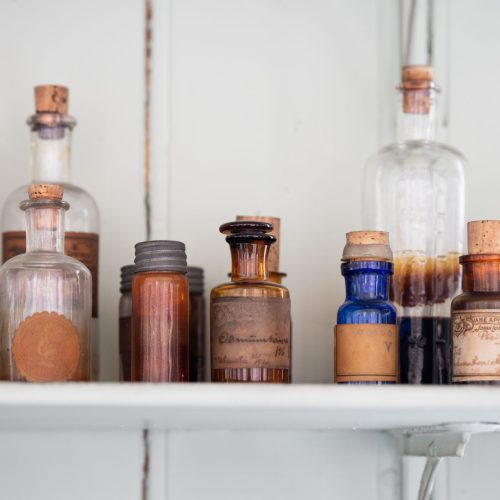
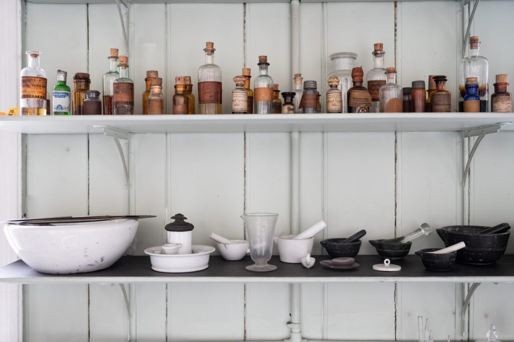
Bottles of dyes – crystal violet, fuchsine and methyl violet
When Armauer Hansen studied tissue samples through his microscope, they were either unstained or stained with osmic acid. Osmic acid stained the entire sample brown with different intensity. This stain did not particularly enhance the visibility of the samples and moreover, they eventually became so dark that they became unusable. Hansen thus had difficulty showing his discovery to his colleagues and convincing them of the presence of the bacterium.
The German physician Albert Neisser was also unable to see bacteria in Hansen’s samples during his visit in 1879. Hansen gave Neisser many tissue samples, and, on his return to Germany, Neisser tried a new staining method developed by doctor Carl Weigert in 1875. Weigert introduced aniline dyes to stain bacteria. These stains based on aniline, a substance distilled from coal tar, were originally developed to dye textiles.
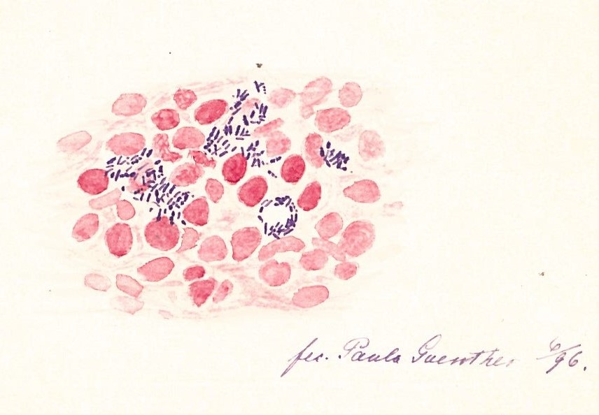
Neisser succeeded in staining the bacterium purple using the dye crystal violet, and the surrounding tissue red using fuchsine. This made the bacteria very visible. Neisser immediately announced his findings at a lecture and published them in a local medical journal. Hansen himself soon succeeded in staining the bacterium with methyl violet, which he described in his 1880 article.
In 1884, the Danish bacteriologist Hans Christian Gram further developed this staining method in order to identify and classify certain types of bacteria. Differentiation of bacteria into gram-positive bacteria, stained purple by crystal violet, and gram-negative bacteria, stained red by fuchsine, still applies today.