Identifying an infectious agent
Even before he was employed as a physician at the hospitals Pleiestiftelsen and Lungegård in 1868, Hansen was convinced that the disease was caused by an infectious agent. If that was the case, it should be detectable in diseased organisms. The question remained, however, of where exactly in the patient the infectious agent could be found, and what kind of infectious agent was he looking for? At that time, many people believed that fungi were the cause of the disease, and that was consequently the first thing Hansen tried to find. The blood samples were kept in a humid container and examined daily for four weeks, with no result.
Could the infectious agent be a bacteria? Bacteriology was still in its infancy, and Hansen had so far only observed and examined putrefying bacteria. Never before had a bacteria been the known cause of a chronic illness. The notion of disease-causing bacteria had just been established.
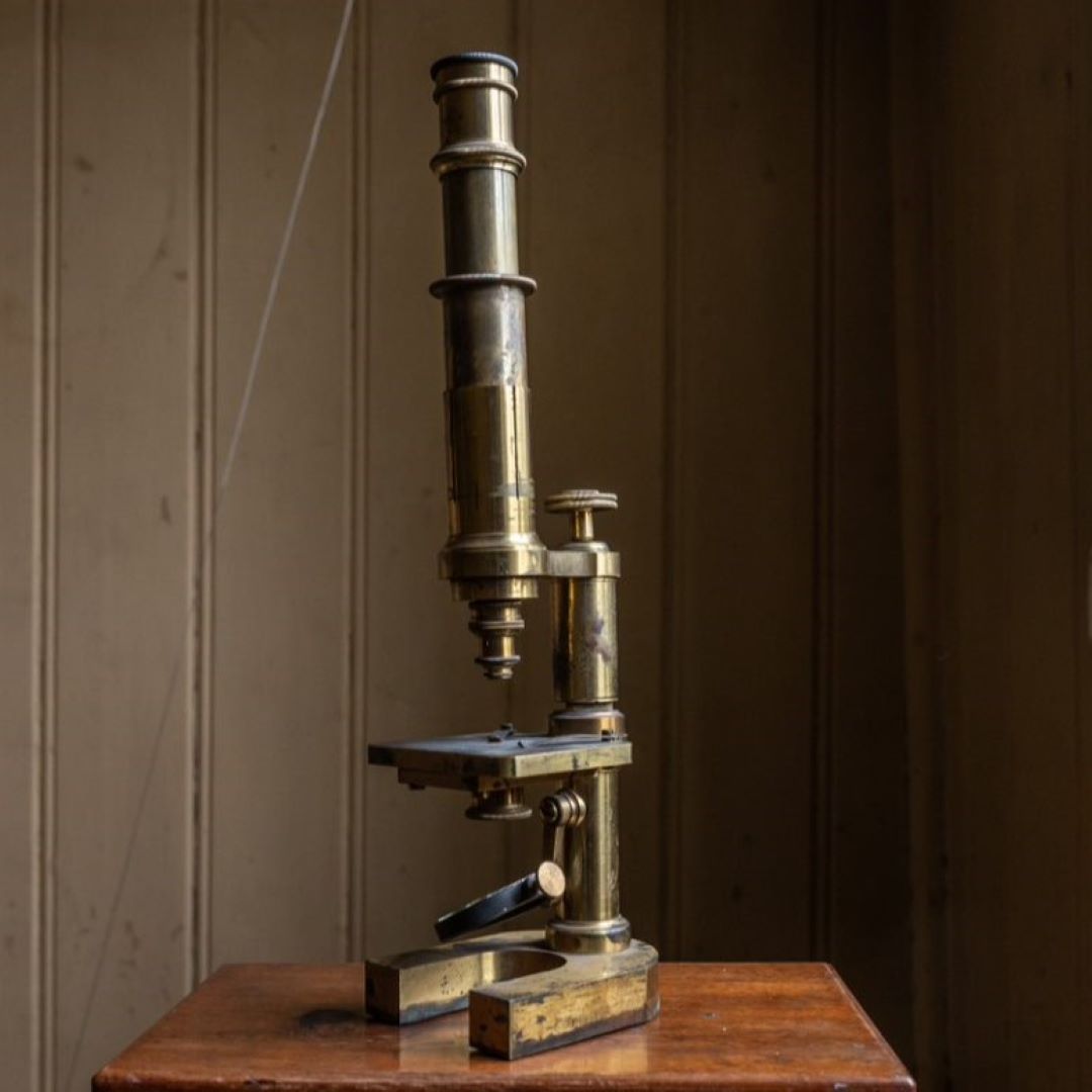
However, bacteria are found everywhere, so how could one rule out bacteria that did not cause disease? First of all, the glass slides on which bacteria were placed had to be free from contamination. Hansen cleaned them with alcohol and burned off the residue. The distilled water he used during the microscopy had to be thoroughly examined. The samples taken from leprosy nodules had to be taken either from excreted fluid or scrapings of undamaged skin – damaged epidermis could contain many other bacteria. And what exactly would these bacteria look like?
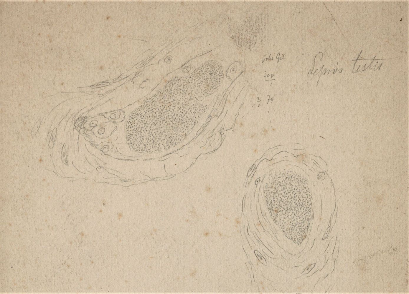
One February evening in 1873, after many long, fruitless days at the microscope, Hansen observed a number of rod-shaped bodies. From Hansen’s notes we can read:
- ‘rod- shaped bodies, either at rest or in slight oscillatory motion’
- ‘if a drop of water is added to the sample, the rods engage in more lively motion, and little by little, more and more rods emerge, the older the nodule, the more there are’
- ‘in some cases, the rods can be seen together in bundles, crossing each other at very sharp angles’
- ‘their size varies greatly, between 0.006-0.0015 mm’
Bergen City Archives.